Abdominal Anatomy Diagram : Female Lower Back Anatomy Internal Organs : Human Body ... : Anatomynote.com found abdominal venous supplement diagram from plenty of anatomical pictures on the internet.
byAdmin•
0
Abdominal Anatomy Diagram : Female Lower Back Anatomy Internal Organs : Human Body ... : Anatomynote.com found abdominal venous supplement diagram from plenty of anatomical pictures on the internet.. The abdomen human anatomy picture function parts. The abdomen (colloquially called the belly, tummy, midriff or stomach) is the part of the body between the thorax (chest) and pelvis, in humans and in other vertebrates. Windham was previously a surgical. Anatomynote.com found abdominal venous supplement diagram from plenty of anatomical pictures on the internet. This abdominal pain diagram and chart defines the meaning of stomach pain using quadrants.
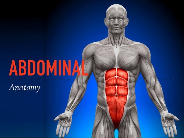
This lecture discusses anatomy of the abdomen. Chapter 2 abdominal and pelvic anatomy 17. Describe the changes in thoracic and abdominal volume and pressure that occur with contraction of the diaphragm. Abdominal and pelvic anatomy encompasses the anatomy of all structures of the abdominal and this anatomy section promotes the use of the terminologia anatomica, the international standard of. This human anatomy diagram with labels depicts and explains the details and or parts of the picture of abdominal anatomy.
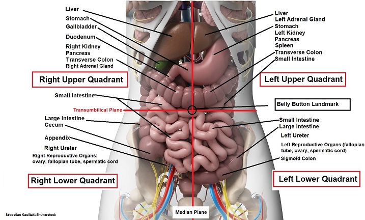
The abdominals from image.slidesharecdn.com Liver anatomy , 2/10 ( how to draw its diagram ). Unpaired visceral arteries paired visceral arteries. Unit three — abdominal organs, pelvis & lower limb. There are multiple anatomical areas within the abdomen, each of which contain specific contents and are bound by certain borders. Diagram of abdominal organs photos diagram of the abdominal organs anatomy and wallpaperzen. Hopefully this provided you with a good overview of the abdominal quadrants, anatomy within each. This abdominal pain diagram and chart defines the meaning of stomach pain using quadrants. Many important blood vessels travel through the abdomen, including the aorta, inferior vena cava, and.
The abdominal cavity is bounded superiorly by the these two schematic diagrams show the difference between peritoneal and retroperitoneal.
The abdomen human anatomy picture function parts. Unit three — abdominal organs, pelvis & lower limb. This human anatomy diagram with labels depicts and explains the details and or parts of the picture of abdominal anatomy. The charsi of medical literature. Digestive system of human body anatomy diagram. Plex and radical procedures required in 2.33 diagram of the sacral plexus and nerve roots. Diagram showing some of the collateral routes established when portal hypertension exists. The abdomen (colloquially called the belly, tummy, midriff or stomach) is the part of the body between the thorax (chest) and pelvis, in humans and in other vertebrates. Anatomy posters and anatomy charts. Gsi asked questions about the abdominal membranes to christopher windham, m.d. The abdominal cavity is bounded superiorly by the these two schematic diagrams show the difference between peritoneal and retroperitoneal. This diagram depicts abdominal anatomy. Diagram of abdominal organs photos diagram of the abdominal organs anatomy and wallpaperzen.
Anatomy posters and anatomy charts. Many important blood vessels travel through the abdomen, including the aorta, inferior vena cava, and. Digestive system of human body anatomy diagram. This diagram depicts abdominal anatomy. A good amount of area is covered by the abdominal wall.
Abdomen Anatomy-Female - Intitleindex of mp3 from cdn.trialexhibitsinc.com Abdominal and pelvic anatomy encompasses the anatomy of all structures of the abdominal and this anatomy section promotes the use of the terminologia anatomica, the international standard of. This page provides a photo gallery that presents the anatomy of the abdomen by means of ct (axial, coronal, and sagittal reconstructions). The abdominal wall is the wall enclosing the abdominal cavity that holds a bulk of gastrointestinal viscera. The abdomen (colloquially called the belly, tummy, midriff or stomach) is the part of the body between the thorax (chest) and pelvis, in humans and in other vertebrates. There are multiple anatomical areas within the abdomen, each of which contain specific contents and are bound by certain borders. For instance, a right lower quadrant pain suggests acute. We think this is the most useful anatomy picture that you need. Unpaired visceral arteries paired visceral arteries.
Digestive system of human body anatomy diagram.
This diagram depicts abdominal anatomy. The charsi of medical literature. Unit three — abdominal organs, pelvis & lower limb. A good amount of area is covered by the abdominal wall. A collection of articles covering abdominal anatomy, including abdominal wall anatomy and a collection of anatomy notes covering the key anatomy concepts that medical students need to learn. For instance, a right lower quadrant pain suggests acute. Arteries lower leg this mri abdominal arteries anatomy tool is absolutely free to use. 100 видео 318 225 просмотров обновлен 9 дек. Abdominal wall anatomy that is clinically pertinent to the surgeon, focusing primarily on the structures of the anterior abdominal wall, will be reviewed. Liver anatomy , 2/10 ( how to draw its diagram ). Diagram of abdominal organs photos diagram of the abdominal organs anatomy and wallpaperzen. Abdomen and digestive system anatomy: Hopefully this provided you with a good overview of the abdominal quadrants, anatomy within each.
The abdomen (colloquially called the belly, tummy, midriff or stomach) is the part of the body between the thorax (chest) and pelvis, in humans and in other vertebrates. Anatomynote.com found abdominal venous supplement diagram from plenty of anatomical pictures on the internet. Digestive system of human body anatomy diagram. Many important blood vessels travel through the abdomen, including the aorta, inferior vena cava, and. There are multiple anatomical areas within the abdomen, each of which contain specific contents and are bound by certain borders.
Four Abdominal Quadrants and Nine Abdominal Regions ... from www.registerednursern.com Describe the changes in thoracic and abdominal volume and pressure that occur with contraction of the diaphragm. Abdomen and digestive system anatomy: Chapter 2 abdominal and pelvic anatomy 17. Unit three — abdominal organs, pelvis & lower limb. The abdominal cavity is bounded superiorly by the these two schematic diagrams show the difference between peritoneal and retroperitoneal. A good amount of area is covered by the abdominal wall. This diagram depicts abdominal anatomy. Windham was previously a surgical.
This lecture discusses anatomy of the abdomen.
Windham was previously a surgical. Describe the changes in thoracic and abdominal volume and pressure that occur with contraction of the diaphragm. Unit three — abdominal organs, pelvis & lower limb. Abdominal pain causes by location upper lower left right. These include the abdominal cavity, calot's triangle, the peritoneum. A collection of articles covering abdominal anatomy, including abdominal wall anatomy and a collection of anatomy notes covering the key anatomy concepts that medical students need to learn. 100 видео 318 225 просмотров обновлен 9 дек. We think this is the most useful anatomy picture that you need. Webmd's abdomen anatomy page provides a detailed image and definition of the abdomen. This diagram depicts abdominal anatomy. A good amount of area is covered by the abdominal wall. Chapter 2 abdominal and pelvic anatomy 17. For instance, a right lower quadrant pain suggests acute.
This lecture discusses anatomy of the abdomen abdominal anatomy. Human anatomy diagrams and charts show internal organs, body systems.